Why Choose a Spinal Interbody Cage for Spinal Fusion?
When a damaged disc causes persistent pain, instability, or nerve pressure, spinal fusion may become a carefully considered option. A Spinal Interbody Cage is placed between two vertebrae after disc removal. It helps restore disc height, support alignment, and create space for bone growth. Imagine a small structural bridge inside the spine. Its role is precise, but not magical.
Spine surgeon Dr. Alexander R. Vaccaro has emphasized, “The goal of spinal fusion is to stabilize the spine while preserving or improving function.” This principle explains why cage selection requires more than choosing a familiar product. Surgeons assess the patient’s diagnosis, bone quality, spinal level, surgical approach, and expected loading forces. Cage material, shape, height, and surface design also matter.
The evidence is encouraging, yet it is not perfect. Fusion can fail. Nerves may remain irritated. Adjacent spinal levels can later develop problems. These possibilities deserve honest discussion. A cage cannot correct every source of back pain. That is an uncomfortable truth.
Clinical experience suggests that the best choice balances stability, biological healing, and safe placement. A surgeon may choose a titanium, polymer, or composite device based on the patient’s anatomy and imaging. The decision should be individualized, documented, and reviewed with realistic expectations. A Spinal Interbody Cage is a tool, not a guarantee. Its value depends on accurate diagnosis, sound technique, and careful follow-up.
What Is a Spinal Interbody Cage?
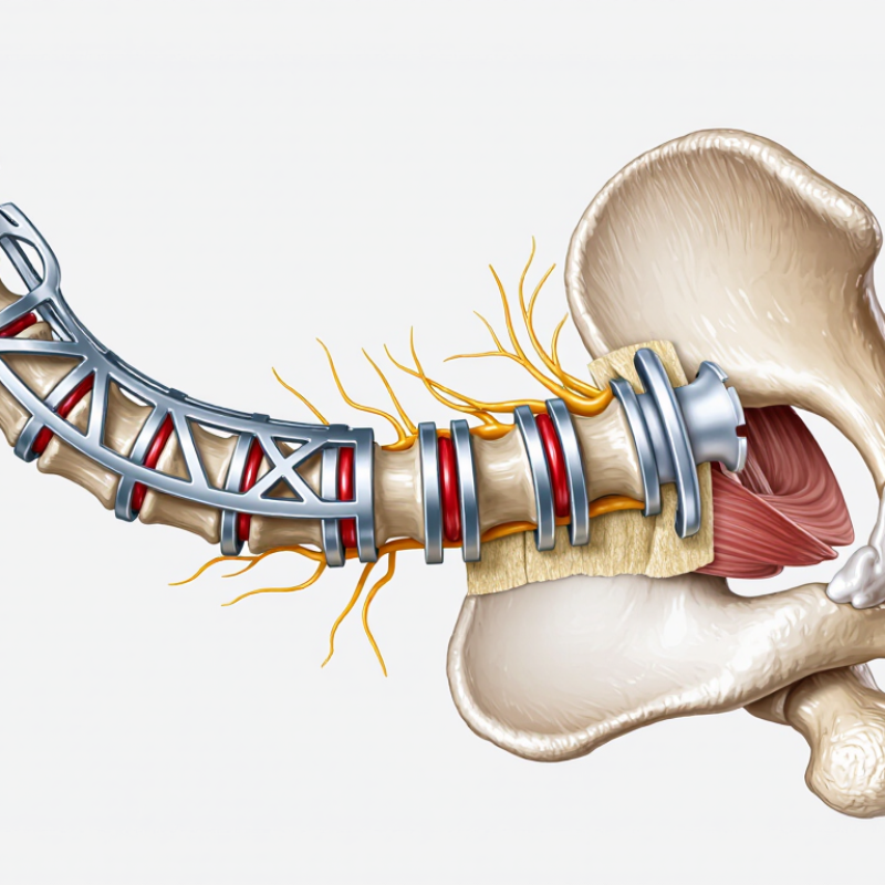
A spinal interbody cage is a small implant placed between two vertebrae during spinal fusion surgery. It replaces a damaged disc and helps restore space between the bones. This space can reduce pressure on nearby nerves. It may also support the spine’s natural alignment.
The cage usually contains a hollow center. Surgeons fill this space with bone graft material, which encourages the vertebrae to grow together. Over time, new bone can form around and through the cage. The implant may be made from medical-grade metal or specialized polymer materials. Its shape and size depend on the patient’s anatomy, the affected spinal level, and the surgical approach.
A cage is not a replacement for careful diagnosis. It does not repair every cause of back pain. Nerve compression, instability, bone quality, and previous surgery can change the decision. Surgeons may also use plates, screws, or rods to improve stability. The procedure carries possible risks, including infection, nerve irritation, nonunion, and persistent pain. Healing can be slow.
The idea seems simple.
In practice, it requires precise planning. Imaging studies help the surgical team measure disc height and evaluate bone structure. A qualified spine specialist should explain why a cage may help, what alternatives exist, and how recovery could affect daily activities. Even technically successful fusion may not remove all discomfort, which is an important detail patients sometimes underestimate.
Why Choose a Spinal Interbody Cage for Spinal Fusion? - What Is a Spinal Interbody Cage?
| Data Dimension | Key Point | Evidence-Based Description |
|---|---|---|
| Definition | A structural implant placed between vertebral bodies | A spinal interbody cage is a hollow or porous device inserted into the disc space after removal of damaged disc material. It helps maintain the space between adjacent vertebrae while bone fusion develops. |
| Primary purpose | Support fusion and restore disc-space height | The cage helps preserve foraminal height, supports the front or central portion of the spinal column, and provides a contained space for bone graft material. |
| How fusion occurs | Bone grows through or around the cage | The cage itself does not create a solid fusion. Bone graft placed inside or around it must bridge the treated vertebrae, gradually forming a stable bone connection. |
| Common indications | Selected degenerative and structural spinal conditions | Potential indications include symptomatic disc degeneration, recurrent disc disease, certain spinal stenosis cases, vertebral instability, spondylolisthesis, deformity, and selected fractures. Suitability depends on imaging, symptoms, bone quality, and overall health. |
| Common surgical approaches | Anterior, posterior, lateral, or oblique access | Cages may be inserted through approaches such as anterior lumbar interbody fusion, posterior or transforaminal lumbar interbody fusion, lateral lumbar interbody fusion, or cervical interbody fusion. The approach is selected according to spinal level, anatomy, and surgical goals. |
| Typical materials | Radiolucent polymers and metal-based materials | Common materials include medical-grade polyetheretherketone, titanium alloys, porous titanium, and selected ceramic or composite materials. Material selection involves considerations such as strength, imaging visibility, bone-contact characteristics, and surgeon preference. |
| Bone graft options | Autograft, allograft, or bone-growth products | Graft material may come from the patient, a screened donor source, or a regulated bone-growth product. The choice depends on the procedure, patient factors, local regulations, and the surgeon’s clinical judgment. |
| Mechanical function | Restores alignment and helps distribute load | A properly selected cage can help restore disc height, improve segmental alignment, and transfer compressive loads between vertebral bodies. Additional fixation may be used to improve immediate stability. |
| Imaging considerations | Some materials make postoperative assessment easier | Radiolucent materials can reduce artifact on radiographs and computed tomography, allowing clearer assessment of bone formation. Metal implants may create more imaging artifact, although modern designs can limit its effect. |
| Potential benefits | Stability, decompression support, and restoration of anatomy | When appropriately indicated, interbody fusion may help maintain disc-space height, support nerve decompression, stabilize painful motion, correct selected alignment problems, and create conditions for solid fusion. |
| Limitations | Fusion is not guaranteed | A cage cannot correct every source of back or neck pain and does not eliminate the possibility of nonunion, recurrent symptoms, adjacent-segment degeneration, or the need for further treatment. |
| Potential complications | Surgical, implant-related, and healing-related risks | Possible complications include infection, bleeding, nerve or vascular injury, dural tear, cage migration or subsidence, nonunion, hardware failure, blood clots, and approach-specific problems. Risk varies by spinal level, surgical approach, bone quality, and patient health. |
| Factors affecting fusion | Patient health and postoperative behavior matter | Smoking or nicotine exposure, poor bone quality, diabetes, obesity, malnutrition, certain medications, multilevel surgery, and inadequate postoperative protection may reduce the likelihood or speed of successful fusion. |
| Recovery and assessment | Healing is gradual and requires follow-up | Pain and activity levels often improve progressively, but bone fusion commonly takes several months and varies between individuals. Follow-up examinations and imaging are used to evaluate alignment, implant position, and evidence of bone bridging. |
| Best candidate principle | Use is based on individualized clinical evaluation | An interbody cage may be considered when symptoms, physical findings, imaging, and non-surgical treatment history support fusion. A qualified spine specialist should determine whether the expected benefits outweigh the individual risks. |
Important: This table provides general educational information and does not replace a diagnosis or treatment plan from a qualified spine specialist.
FAQS
An interbody cage replaces a damaged disc space between two vertebrae. It supports the area after disc removal.
The cage holds bone graft material in a contained space. New bone may grow around or through it, joining the vertebrae.
No. Bone healing creates the fusion. Screws and rods may limit movement while the bone develops.
Restored height may improve spinal alignment and enlarge narrowed nerve passages. It may also reduce pressure near irritated nerves.
Static cages keep one height. Expandable cages adjust inside the disc space. Angled designs can support spinal alignment.
There is no universal best material. Some materials improve imaging, while others provide strong support or bone attachment.
Bone quality, smoking, nutrition, activity, and general health can influence healing. Osteoporosis may increase settling risk.
Avoid lifting too soon, even when pain improves. Keep imaging appointments. Report fever, drainage, weakness, or new numbness promptly.
Not always. Small setbacks can happen. They deserve attention, not panic, although the ideal response is not always obvious.
Conclusion
A Spinal Interbody Cage is a medical device placed between two vertebrae during spinal fusion surgery. By restoring disc height and helping maintain spinal alignment, it creates a stable space where bone graft material can support the growth of new bone. Over time, this process may join the adjacent vertebrae into a solid segment and help reduce motion-related pain or instability. Interbody cages may also help improve foraminal space, potentially reducing pressure on nearby nerves.
These cages are available in different shapes, designs, and materials, such as medical-grade polymers, titanium, or composite structures. The most suitable option depends on the patient’s spinal condition, anatomy, bone quality, surgical approach, and overall health. Although an interbody cage can provide important support, spinal fusion still involves risks, including infection, nerve irritation, nonunion, implant displacement, and continued symptoms. Patients should discuss expected benefits, limitations, alternatives, and recovery requirements with a qualified spinal specialist before making a treatment decision.